Cone Dystrophies
This is a group of clinically heterogeneous diseases which either occur sporadically or as familial traits. In most cases, cone function is normal after birth. The age of onset varies from the late teens into the sixties. Patients experience progressive visual problems including daytime and color vision, as well as visual acuity.
The mode of inheritance can be autosomal dominant, recessive or X linked. Interestingly, mutations in the same gene can lead to retinitis pigmentosa or cone dystrophy, as reported for RPGR mutations.
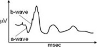
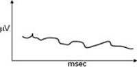
We identified mutations in a regulatory calcium channel subunit in a mouse line with an autosomal recessive retinal disease as well as in two affected siblings with a mild form of autosomal recessive cone dystrophy. The mutant mouse line showed reduced rod responses and completely absent cone activities in electrophysiology (electroretinography, ERG). Morphologically, upon electron microscopy, ribbon synapses were abnormal or absent and the synaptic layer between photoreceptor and bipolar cells (outer plexiform layer) is reduced. We mapped the gene defect and identified a frameshift mutation in the Cacna2d4 gene, which encodes an L-type calcium channel auxiliary subunit of the alpha-2-delta type. This subunit is believed to regulate channel density/number on the cell surface as well as their activity.
Subsequently, mutation screening in patients with the initial diagnosis of night blindness revealed a premature stop codon in exon 25 (out of 38) of CACNA2D4. However, both patients show symptoms of a slowly progressing cone dystrophy.
Current work is focusing on the identification of the respective alpha-1 subunit, which is the target of CACNA2D4.
TEAM MEMBERS
Wolfgang Berger (PhD)
Silke Feil
Samuel Koller (PhD)
Jordi Maggi (PhD)
COLLABORATORS
Daniel Barthelmes (MD, PhD), Dept. Ophthalmology, Zurich
Christina Gerth-Kahlert (MD), Dept. Ophthalmology, Zurich