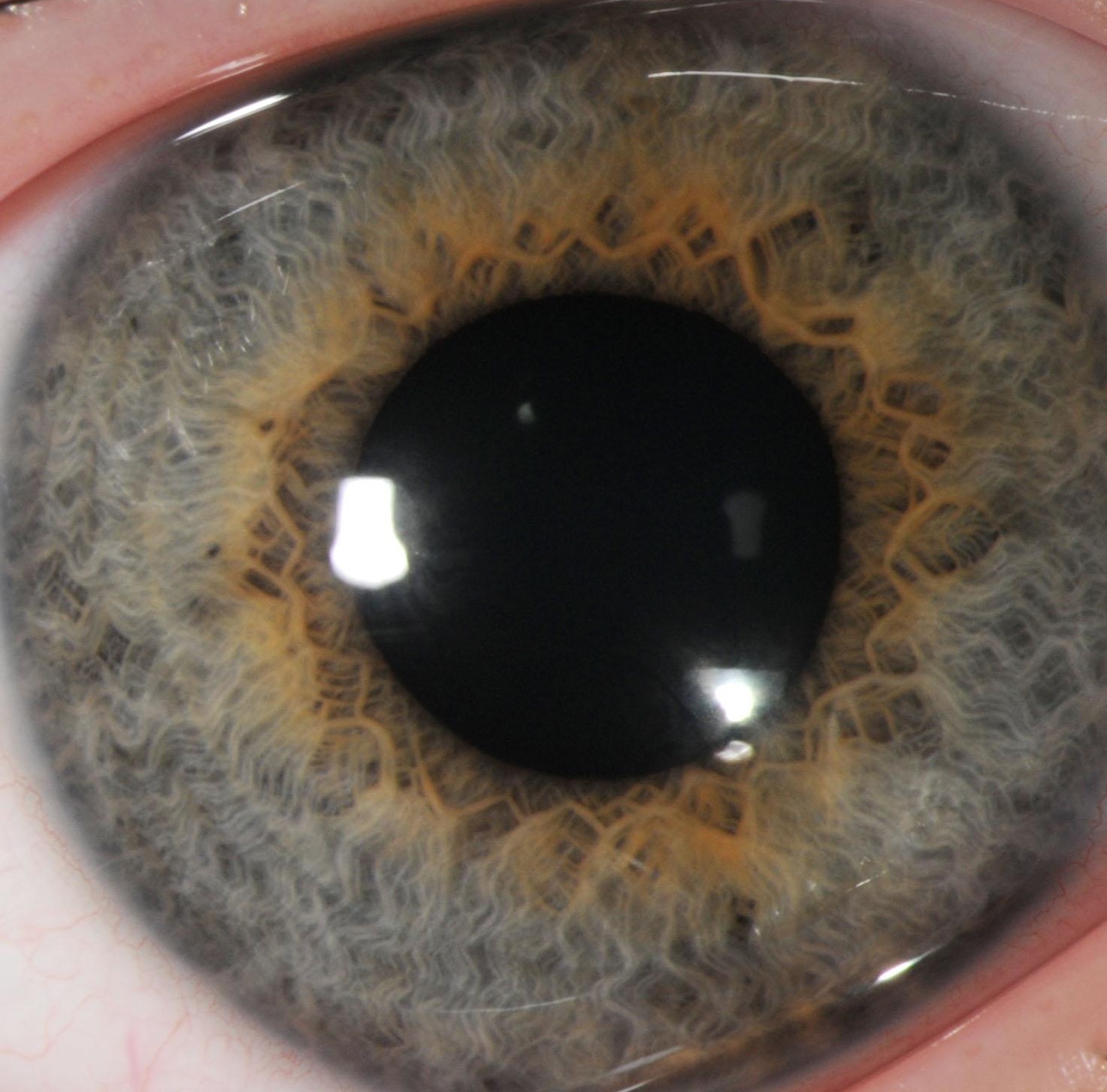
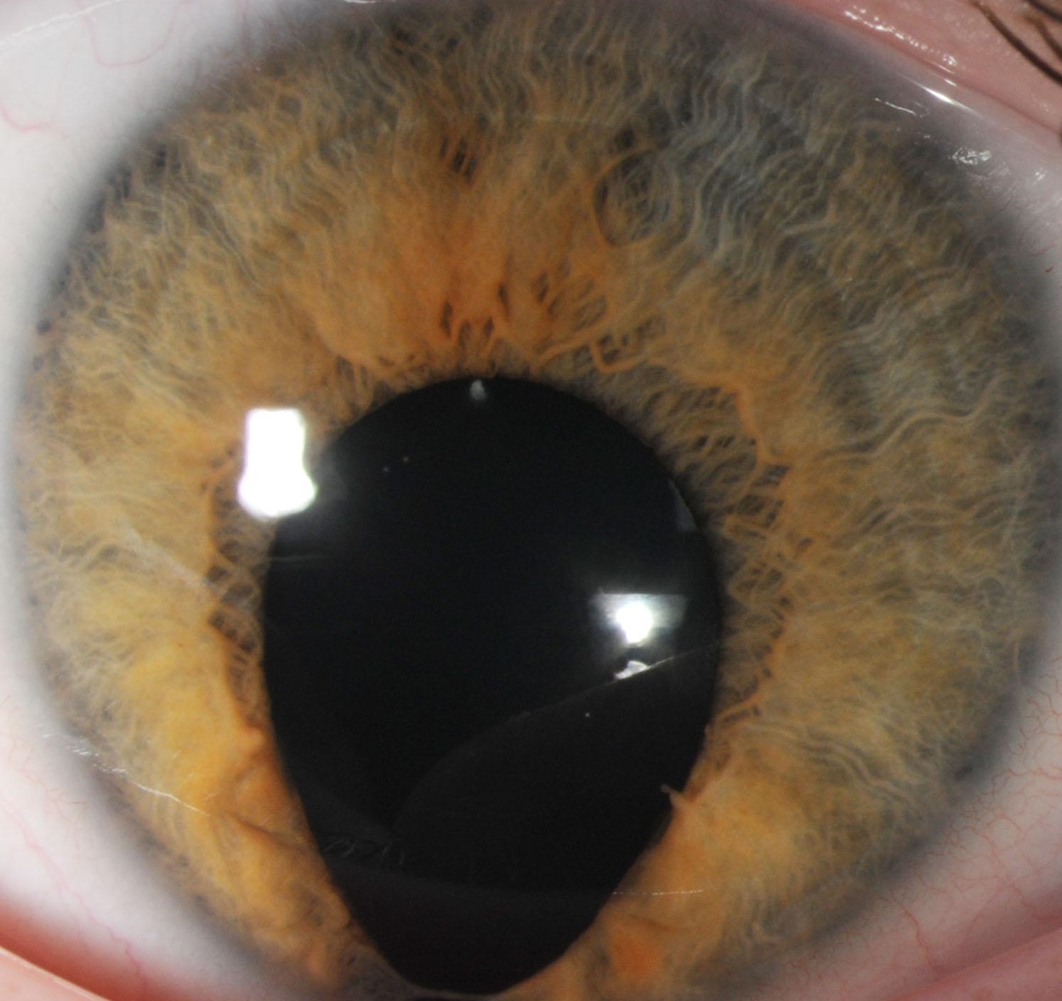
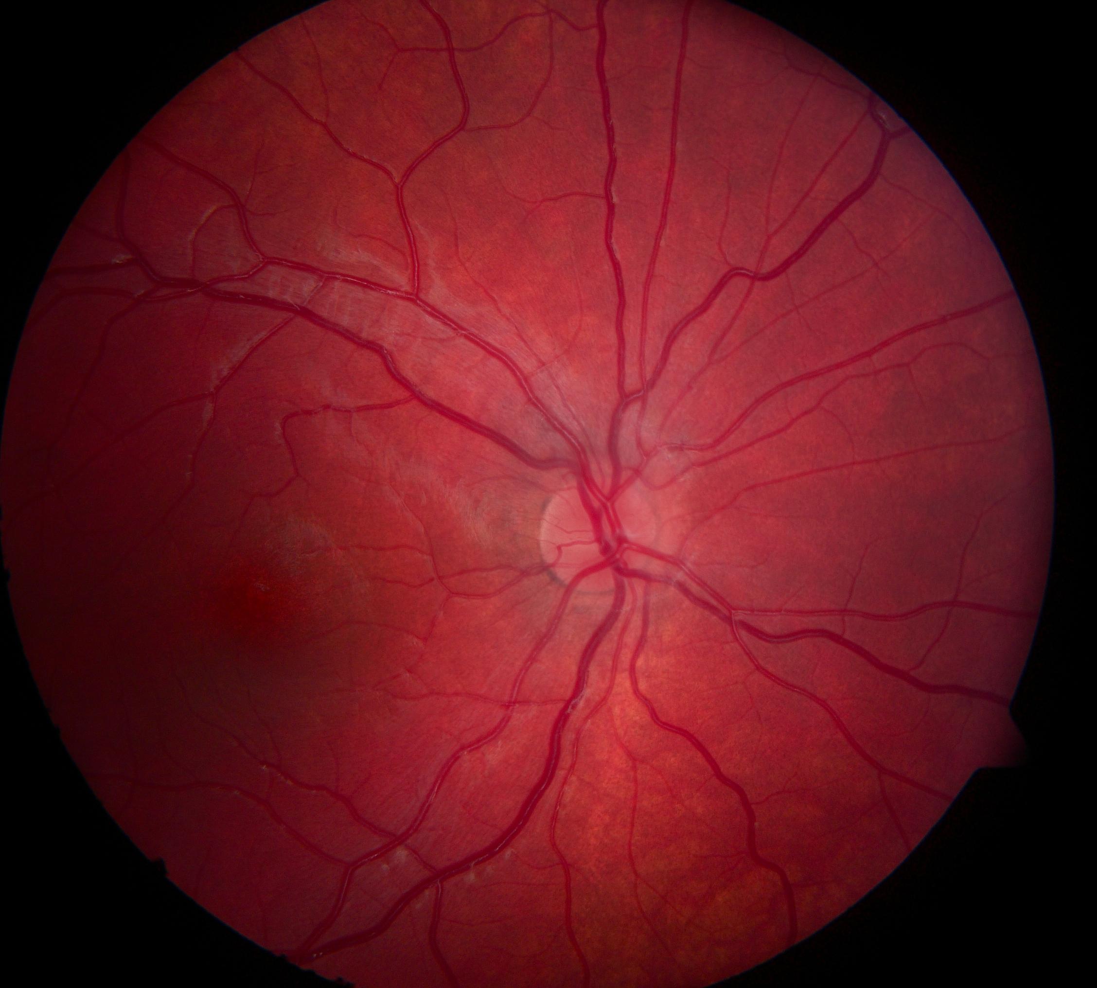
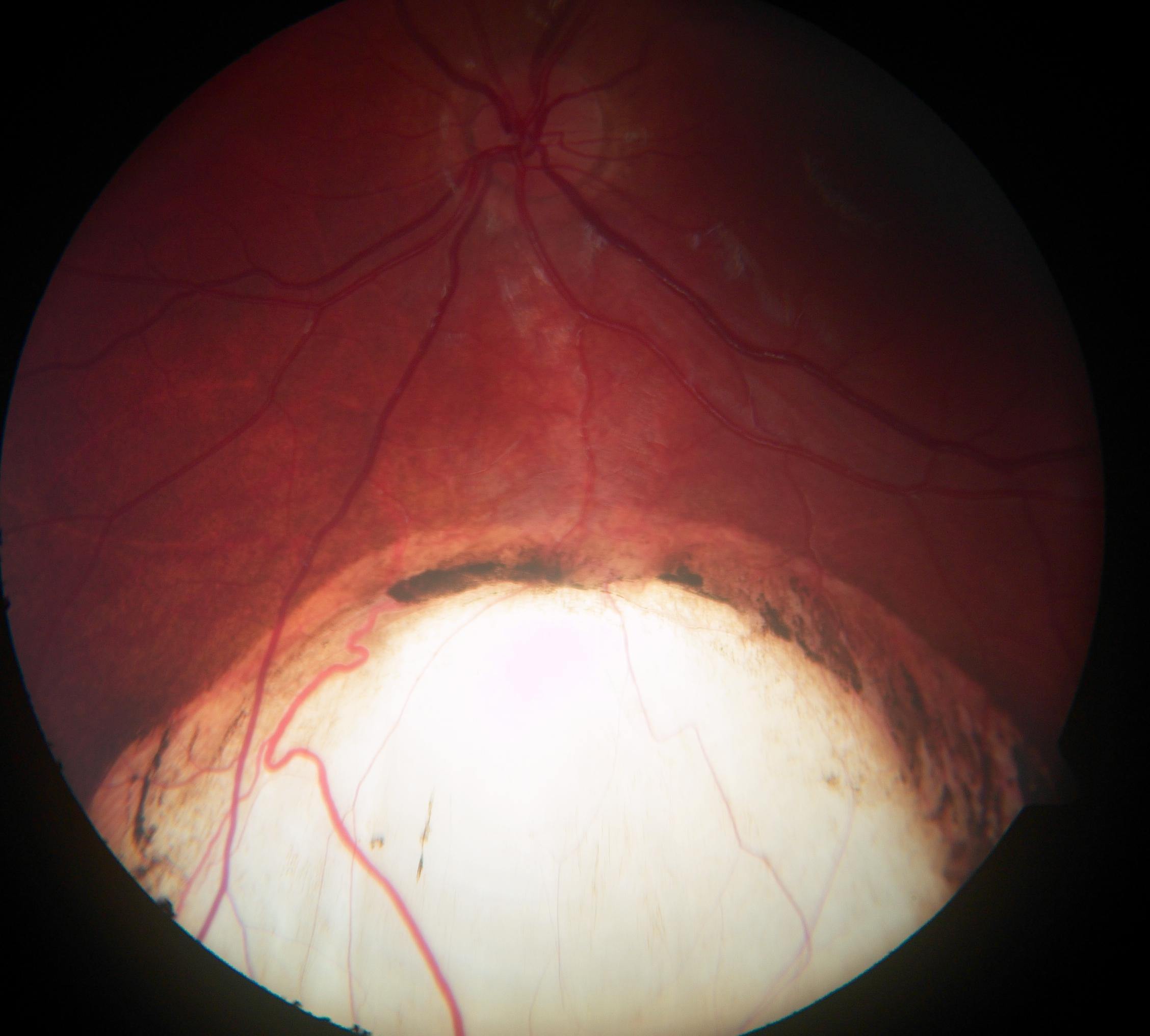
Coloboma and Microphthalmia
Ocular coloboma and microphthalmia (C/M) are related congenital eye malformations and refer to a segmental defect affecting all or parts of the iris, lens, choroid, retina, and optic nerve. It is typically caused by a partial or complete failure of optic fissure closure during early eye development.
Microphthalmia is characterized by a reduced axial length of the eye. It primarily results from reduced growth of the eye but can also occur secondary to incomplete optic fissure closure. However, the exact pathophysiological mechanisms responsible for microphthalmia are still poorly understood. It remains unknown whether the optic fissure defects in colobomatous microphthalmia are the cause or consequence of ocular growth defects.
C/M can be associated with severely reduced visual development or deterioration of vision through complications such as cataract or retinal detachment and accounts for approximately 15% of severe visual impairment and blindness worldwide. The prevalence is estimated to range from 2 to 17 per 100,000 births for microphthalmia and from 2 to 14 per 100,000 births for ocular coloboma. The diseases are genetically heterogeneous with reduced penetrance and variable expressivity even within the same family and between the eyes of the same patient. They may occur isolated or as part of a syndrome.
We performed genetic analysis, including copy number analysis (CNVs), in 19 patients from 15 unrelated families with coloboma and/or microphthalmia using whole exome sequencing (WES) and data analysis for 307 genes of interest.
We identified seven novel and one recurrent potentially disease-causing variants in CRIM1, CHD7, FAT1, PTCH1, PUF60, BRPF1, and TGFB2 in 47% of our families, three of which occurred de novo. The detection rate in patients with ocular and extraocular manifestations (67%) was higher than in patients with an isolated ocular phenotype (46%). Our study highlights the significant genetic heterogeneity in C/M cohorts and emphasizes the diagnostic power of WES for the screening of patients and families with C/M.
TEAM MEMBERS
Wolfgang Berger (PhD)
Silke Feil
Patricia Haug
Samuel Koller (PhD)
Elena Lang (MD)
Jordi Maggi (PhD)
Agnès Wlodarczyk
COLLABORATORS
Christina Gerth-Kahlert (MD), Dept. Ophthalmology, University Hospital Zurich